Branch Retinal Artery Occulsion

About Branch Retinal Artery Occlusion

A branch retinal artery occlusion (BRAO) is usually marked by a painless and abrupt onset of peripheral vision loss and, in some cases, central vision loss. The cause of the condition is usually related to an embolus (clot or plaque) from a carotid artery (in the neck) or the heart. While no ocular therapy has been proven valuable, some ophthalmologists may attempt to dislodge the embolus if the condition has been present for less than 24 hours. Methods include ocular massage, anterior chamber paracentesis (tapping fluid from the eye), or administration of glaucoma agents.

Treatment

01-brao-fa_420.jpg

Loss of visual acuity with a BRAO will mostly depend on whether there is disruption of arterial blood flow to the macula or the presence of significant macular edema (swelling). Patients will be evaluated for cardiovascular risk factors and treated accordingly, often with a family or internal medicine physician involved. The majority of patients are found to have carotid artery disease (narrowing), high blood pressure, cardiac disease, or combinations of these disorders. Less commonly, infectious, inflammatory or hereditary conditions can be the cause.

Most patients will be evaluated from an ophthalmic standpoint every 3 to 6 months until stable. The prognosis for vision depends on whether or not the central macula was initially affected. In general, poor vision upon presentation is indicative of a poor visual outcome. Approximately 90% of patients will recover visual acuity of 20/40 or better, though most patients will have noticeable permanent visual deficits.

In rare cases patients may develop other complications as a result of BRAO, such as neovascularization of the retina and iris and neovascular glaucoma. Now, new treatments are available which can preserve and even restore vision in some patients. We specialize in these therapies here at the Retina Macula Institute.

Central Serous Choroidopathy

What is Central Serous Choroidopathy?

central-serous-choroidopathy_4202.jpg

Central Serous Choroidopathy (CSC), also known as Central Serous Retinopathy (CSR), is a disease that causes fluid build-up beneath the retina. This leakage of fluid comes from the layers under the retina, the retinal pigment epithelium (RPE) and choroid. This condition generally affects a person’s central vision.

Diagnosis

Central serous can be diagnosed by examining the retina with special lenses allowing a view of the back of the eye. A blistering of the retina may be seen. Some cases are very subtle and additional testing is required. Fluorescein angiography will show a spot of leakage which, in some cases, might have accumulated into a classic “smoke-stack” pattern and may be used to identify complications including choroidal neovascularization. Indocyanine Green (ICG) angiography supports the role of the choroid in this disease and is rarely useful in management. We now use high-resolution optical coherence tomography (OCT) to identify fluid under the retina and to identify subtle cases that might otherwise be missed.

Who is at risk?

While there is no definite known cause for this condition, we do know that it usually affects young males between the ages of 20-45. However, women may also be affected. Risk factors for the development of this condition include a “type A personality”, the use of steroids, caffeine or other stimulants (e.g., energy drinks, dietary supplements), as well as smoking, a history of trauma, physical/emotional stress.

Signs and Symptoms

Signs of central serous can include an elevation of the central retina (macula), deposits beneath the retina, dark and light color changes (hyper- and hypo-pigmentation), and, in complex cases, blood, fat deposits (lipid) and scarring (fibrosis). The “normal eye” often has subtle changes as well. Symptoms include blurred central vision, small image size (micropsia), and distortion of the image (metamorphosia). After central serous resolves, most patients will still have residual symptoms such as distortion, decreased color and contrast sensitivity, and vision difficulty at night. There is also a 40-50% change of recurrence over time. In some cases, the longterm complications may be due to residual chronic activity and a more detailed evaluation of the disease process may improve the visual results.

Treatments

While there is no known cure, treatment is directed at the underlying modifiable risk factors such as removal of stimulants from the diet (e.g., smoking, caffeine, energy drinks, steroids) and improved life style (e.g., more sleep = less stress). Normally this condition clears up in a month or two. In patients with more severe vision loss or a prolonged course of the disease, laser treatment has been shown to reduce the course of the disease and improve vision. In some complex cases, we may consider treatment with a “cold laser” (photodynamic therapy with Visudyne), intraocular injections of anti-VEGF drugs such as Avastin, or a combination of these treatments. Non-steroidal anti-inflammatory drugs (NSAIDS) have been shown, experimentally, to stimulate fluid absorption but have not been proven in a controlled clinical trial. The newest treatment options may improve outcomes and we specialize in the latest testing and treatments here at the Retina Macula Institute.


Cytomegalovirus (CMV) Retinitis

What is Cytomegalovirus (CMV) Retinitis?

CMV_400.jpg

Cytomegalovirus (CMV) retinitis is a sight-threatening disease associated with late-stage AIDS. In the past, about one-fourth of active AIDS (Acquired Immunodeficiency Syndrome) patients developed CMV retinitis. However, this figure appears to be dropping thanks to a potent combination of drugs that help restore the function of the immune system.

When CMV invades the retina, it begins to compromise the light-sensitive receptors that enable us to see. This does not cause any pain, but you may see floaters or small specks and experience decreased visual acuity (blurry vision) or decreased peripheral vision. Light flashes and sudden loss of vision can also occur.

The disease usually starts in one eye but often involves both eyes. If left untreated, CMV retinitis can cause retinal detachment and blindness within just two to six months. AIDS patients sometimes also experience changes to the retina and optic nerve without clear signs of CMV retinitis. CMV retinitis is caused by the common cytomegalovirus; nearly 80% of adults harbor antibodies to CMV, which indicate that their bodies have fought the virus off successfully.

In the case of AIDS, patients’ weakened or non-functioning immune systems are unable to stave off this virus. Other people with a weakened or suppressed immune system, such as those undergoing chemotherapy or a bone marrow transplant, are also at risk. Newer treatments are now available that can restore vision in some patients with severe uveitis and we specialize in these therapies here at the Retina Macula Institute.


Diabetic Retinopathy

What is Diabetic Retinopathy?

diabetic-retinopathy_420.jpg

Diabetic Retinopathy is a disease of the retina. The retina is the nerve layer that lines the back of your eye. It is the part of your eye that “takes pictures” and sends the images to your brain. Many people with diabetes get retinopathy. This kind of retinopathy is called diabetic retinopathy (retinal disease caused by diabetes).

Diagnosis

An eye exam by an eye specialist (ophthalmologist or optometrist) is the only way to diagnose diabetic retinopathy. Having an eye exam every year can help find retinopathy before it changes your vision. Specialized testing such as fluorescein angiography, optical coherence tomography and ultrasonography may be required to determine how bad the eye is affected and to select the best treatment options.

Who is at risk?

Every patient with diabetes is at risk for developing diabetic retinopathy. The higher the blood sugar, the faster you will develop the disease. While most are diabetic for 10 years before the retina is affected, some develop it after just a few years or months. Diabetic retinopathy happens when high blood sugar damages the tiny blood vessels of the retina. When you have diabetic retinopathy, high blood pressure can make it worse. High blood pressure can cause more damage to the weakened vessels in your eye, clouding more of your vision. High levels of fat in the blood, smoking, and obesity are also risk factors as are certain medications.

Signs and Symptoms

The signs of diabetic retinopathy (DR) include blood spots, fluid (edema) and abnormal blood vessel growth. Fluid threatening the center of vision is termed clinically signifcant diabetic macular edema (CSME) and requires treatment. Poor circualtion from damaged blood vessels leads to abnormal blood vessel growth termed proliferative diabetic retinopathy and usually requires treatment as well. The symptoms range from no change in vision to some blurring of vision with macular edema to black dots floating in the vision and mild to severe, even complete vision loss. Don’t wait for symptoms. Be sure to have a comprehensive dilated eye exam at least once a year.

Treatment

You may not need treatment for diabetic retinopathy when the changes are early. In more advanced cases, surgery, laser treatment, or medicines may help slow the vision loss caused by diabetic retinopathy. You may need to be treated more than once as the disease gets worse. The goal of treatment is to reduce the rate of vision loss or to stabilize vision. In select patients, such as those with severe swelling of the retina or blood filling the inside of the eye, dramatic improvement in vision may be possible.

Unfortunately, Diabetic retinopathy remains the leading cause of blindness in the working age, and even with excellent care, further vision loss may occur. However, newer treatments are now available that have been known to restore vision in some patients. We specialize in these therapies here at the Retina Macula Institute.

Dislocated IOL

What is Dislocated IOL?

pseudophakia_420.jpg

An intraocular lens implant is usually placed in the eye after cataract surgery and may occasionally become malpositioned or dislocated. This may occur as the result of trauma at the time of surgery, previous trauma to the eye that damaged the natural lens or becasue of a systemic disease. Vision may be blurry, double and reduced to the point of legal blindess. Most cases can be readily treated with surgery.

Diagnosis

A dislocated IOL can be identified by examining the front of the eye with a microscope (slit lamp biomicroscopy) as well as the back of the eye using other lenses for viewing the retina. The lens may be falling down from its normal position (sunset syndrome) or completed displaced into the back of the eye.

Signs and Symptoms

Signs of a dislocated intraocular lens (IOL) are the following: the absence of the lens when examining the front part of the eye, viewing the edge of the lens in the slit lamp or seeing the lens at the back of the eye. The patient may notice the edge of the lens implant or even have double vision. If the lens implant dislocates entirely out of the visual axis, the patient may have a very substantial drop in visual acuity, usually to the “counting fingers” level.

Who is at risk?

Patients at increased risk for a dislocated IOL may have a medical condition such as pseudoexfoliation, Marfan’s Syndrome or homocystinuria. In each of these cases the fibers that hold the natural lens in place are defective due to to an abnormal protein made by the body. Fibers that contain the protein normally hold the lens in place. In some cases these preexisting conditions may not be diagnosed. Trauma is also a risk factor and cataracts that develop after trauma should be managed more carefully. Finally, the natural lens of the eye includes a very thin capsule that can break during surgery, even in the best of hands. If the lens is not well supported by the capsule it also might also dislocate.

Treatment

Dislocated IOL is treated by moving the lens into the correct position, replaceing the lens or sewing a lens in place. In most cases the gel in the back of the eye, or vitreous, must be removed in part through the surgical process called “vitrectomy”. Once the lens is free of the vitreous gel it can be moved more safely and repositioned or replaced. In some cases a new lens can also be sewn into the back of the eye.


Endophthalmitis

What is Endophthalmitis?

endopthalmitis_400.jpg

Endophtalmitis is an internal eye infection, also known as an intraocular infection. There are many different types of endophthalmitis, but this section will focus on the most common type, called post-operative endophthalmitis. Most commonly, this condition occurs as a result of cataract surgery and afflicts about 1 in every 1,000 patients. The condition is not necessary attributed to poor surgical execution; it may occur even when the procedure is routine and uncomplicated. Of course, surgical complications may increase the incidence of this infection. Symptoms include sudden-onset vision deterioration, pain, and red eye, which usually occurs 3 to 12 days following surgery.

Who is at risk?

The most common cause of endophthalmitis is bad luck; in fact, every individual shares a 1 in 1000 chance of affliction. However, patients with a history of traumatic cataract, diabetes or those who experienced complications during surgery, are at an increased risk of infection. Post-operative endophthalmitis is classified as a bacterial infection, most often attributed to an organism, which can live benignly on the external portions of the eye. The most common bacterial species is a type of “staph” infection known as Staphylococcus epidermidis. Other “staph” and “strep” (streptococcal) species are only slightly less typical. The least common infectious bacteria are known as gram-negative organisms, the worst of which is an organism called Pseudomonas. Infection by this organism may lead to a poor prognosis.

Treatment

Antibiotics and, sometimes, steroids are injected into the eye and the patient is started on frequent topical antibiotic eye-drops. Patients with very poor presenting acuity are sometimes scheduled for a vitrectomy (surgical removal of the vitreous humor) as this can improve the prognosis for that group of patients.

Despite the severity of this surgical complication, recent improvements in management and treatment have promised many patients a more favorable outcome. At the Retina Macula Institute we specialize in the most advanced treatments available, taking an aggressive and timely approach.


Macular Degeneration

What is Macular Degeneration?

macular-degeneration-wet_400.jpg

The macula is a critical part of the eye and is responsible for detailed vision. Although very small, the macula has the highest concentration of cells in the retina. These particular cells are called photoreceptors and are responsible for converting light into electricity. Photoreceptors allow focused vision, necessary for reading, seeing faces, and drive cars. Damage to these cells is the cause of vision loss in macular degeneration. The area of the retina, just outside the macula, is responsible for peripheral vision and is unaffected by macular degeneration. This area has much lower resolution but allows us to see larger objects. Therefore, patients with macular degeneration will never go completely blind. Macular degeneration comes in two forms. In the dry form of macular degeneration, tiny yellow deposits develop beneath the retina, called drusen. Areas of thinning, called atrophy, may also occur. Approximately 10% of patients with dry macular degeneration will experience enough damage to develop the second form of the disease, called wet macular degeneration. In this form, abnormal blood vessels grow beneath the retina in an effort to bring more nutrients to the diseased tissues. However, these abnormal vessels often leak and form scar tissue. This can be the cause of permanent central vision loss if left untreated. Early signs of wet macular degeneration include central vision distortion, characterized by wavy lines and dark or missing areas of images. An Amsler grid is useful for detecting these initial changes. When diagnosed in the early stages and utilizing the latest technologies and treatments, it is possible that vision might be preserved or restored. Therefore, patients with macular degeneration should monitor their eyesight with an Amsler grid, daily, and visit their eye doctor on a regular basis.

Age-Related Macular Degeneration Causes

Macular degeneration is caused by a number of factors including, but not limited to, age, genetics, and increased oxidative stress on the eye. Specific risk factors include advanced age, genetics (heredity), race, smoking, exposure to sunlight, diets high in saturated fats and cholesterol, little exercise, vascular disease, high blood pressure, and farsightedness (hyperopia). While age and family history are unpreventable, one has the ability to reduce some of the other risk factors. It is important to work with your medical doctor and eye doctor to make these changes.

Can macular degeneration be prevented?

There are a number of factors, within your control, that may help reduce the risk of developing macular degeneration or reduce the progression of a diagnosis. Specifically, one should consider protecting themselves with UV light resistant sunglasses, exercise, vitamin supplementation and a healthy diet. Vitamins have been shown to reduce disease progression by up to 25% while diet may reduce progression by up to 50%. Specifically consider supplements that follow the guidelines set by Age-Related Macular Degeneration studies and modify according to your age, disease severity and medical history. If possible, your diet should include the consumption of green leafy vegetables and fish, at least twice a week. Avoid eating red meat or processed baked goods more than once a week and avoid smoking and sleep deprivation. Omega 3 fatty acids should be consumed once a day along with regular exercise, under the direction of your medical doctor.

How is it treated?

Dry macular degeneration is treated with the preventative guidelines noted above. For the treatment of wet macular degeneration there is a host of new options. There are injections available to stop or reverse the growth of the abnormal blood vessel, which causes leakage and vision loss. In some cases, surgery may be recommended. Clinical studies are now underway to assess new treatments. The goal of treatment is to reduce or stabilize vision loss. In select patients, particularly those diagnosed early, dramatic improvement in vision may be possible. We specialize in the newest restorative therapies, here at the Retina Macula Institute.


Macular Hole

What is a Macular Hole?

macular-hole_400.jpg

The macula provides the sharp, central vision necessary for reading, driving, and seeing fine detail. A macular hole is a small break in the macula, located in the center of the eye’s light-sensitive tissue, called the retina. Here at RMI we review who is at risk for this condition, and apply the most advanced diagnoses and sight-saving treatments available today.
Who is at Risk?

In 80% of cases, macular holes are associated with aging, most common in those over the age of 60. Near-sightedness, trauma, systemic diseases and inflammation account for the other 20%. Ten percent of patients will go on to develop the condition bilaterally. Females tend to be affected more commonly than males and there are theories suggesting its link with shifting estrogen levels around the time of menopause.

Signs and Symptoms

Some of the most common symptoms include distortion, reading difficulty, or a blind spot in the center of vision (scotoma). If the non-dominant eye is affected, however, the condition may go undetected until recognized in a routine eye exam or when the good eye is accidentally covered while performing a visual task.
Diagnosis

While an ophthalmoscope is often used to detect a macular issue, additional tests may be required in order to confirm the presence of a macular hole. Such tests include flourescein angiography (FA) and optical coherence tomography (OCT). An FA may be used to identify associated conditions such macular edema from diabetic retinopathy or macular degeneration. An OCT is used to clearly identify a hole and any associated features, including an epiretinal membrane, vitreous traction and macular edema.

Separation of the vitreous gel from the retina leads to a tear in the macula and the development of the hole, which progresses in stages:

  • macular cyst
  • full thickness hole <400 microns in size
  • full thickness hole >400 microns in size
  • full thickness hole of any size associated with complete posterior vitreous detachment (PVD).

Treatment

mac-hole-photo_3001.jpg

A vitrectomy is required for Macular Hole repair. This procedure entails the removal of the vitreous gel that fills the back of the eye and replacing it with a gas bubble. The fine internal limiting membrane (ILM), located around the edge of the hole, is often removed at the same time to increase the procedure’s success rate. These combined procedures are individually termed, as follows: vitrectomy, membrane peeling and fluid-gas exchange. An Intraocular Laser (“Endolaser”) may also be applied if holes, tears or retinal thinning is found at the time of surgery. Patients must remain face down for 4-14 days after surgery to achieve the best possible results. However, the prognosis for visual recovery is dependent on additional factors, such as the duration that the hole existed before surgery and the preexistence of conditions, such as macular degeneration.

The purpose of the gas bubble is to hold the edges of the hole together while the retina heals. A face down positioning device, like a massage table, can be ordered to help patients comply with these instructions. All surgeries have potential risks and benefits as well as alternatives (e.g., observation) which should be discussed with your doctor. At RMI we provide the most advanced care to optimize recovery of vision.


Macular Pucker

What is Macular Pucker?

Macular pucker, also known as an epiretinal membrane or cellophane maculopathy, is the formation of scar tissue over the macula or the central portion of the retina. The retina is the light-sensitive film at the back of the eye, which converts light into an electrical signal that can be interpreted by the brain. The macula is responsible for detailed vision and, thus, functional vision, necessary for reading and driving. Fortunately, a macular pucker is treatable.
Diagnosis

A macular pucker is usually diagnosed through examination of the retina with a microscope and special lenses. When the condition is present, a membrane or film will appear on the retinal surface, distorting retinal vessels and causing the retina to swell with fluid (edema). Additional tests include the use of fluorescein angiography, to determine if there is any associated condition such as a blocked blood vessel (e.g., “vein occlusion”). The angiogram can also reveal leakage from the distorted blood vessels of the retina which, when present, may lead to more rapid and irreversible loss of vision. Optical coherence tomography (OCT) is useful in the identification of the pucker itself (the membrane on the retinal surface), in measuring the amount of edema and in identifying the presence of additional damage to the retina (e.g., lamellar hole), which may limit the prognosis.

Who is at risk?

macular-pucker_420.jpg

The most common cause of macular pucker is the aging process. Aging of the vitreous — the jelly-like substance that fills about 80% of eyes interior and helps to maintain the eye’s shape — initiates the process. As the vitreous ages, it shrinks and pulls away from the surface of the retina causing “traction”. The traction (or pulling) by the vitreous creates cracks in the surface of the retina stimulating a healing response. Part of this healing respons is the growth of scar tissue on the retina surface, forming a membrane. Contraction of the membrane or scar tissue causes the macula to “pucker”. In most patients with macular pucker the vitreous has detached completely (posterior vitreous detachment) and since this occurs as you age (typically after 65) most patients with macular pucker are elderly.

Macular pucker may also occur after intra-ocular surgery – particularly in cases where retinal tears or a retinal detachment has occurred. Indeed, 15% of patients with a macular pucker have a retinal tear already. Other exacerbating factors include internal inflammation, swelling, holes, tears, or trauma. An onset of flashing lights or floaters may be a warning sign that a change is taking place within your eye.

Signs and Symptoms

Signs of macular pucker include blood vessel distortion, due to the presence of scar tissue on the retina. This can be difficult to detect; sometimes, an abnormal light reflex may be the only clue. Symptoms range from nonexistent to severe. There may be blurring of central vision, distortion (metamorphopsia), change in the size of objects, double vision, and/or loss of depth perception.

Treatment

In cases where the affects on vision are minimal, it is unnecessary to treat macular pucker. When the change in vision is mild and retinal edema is present, some patients will respond to anti-inflammatory drops or injections. When the affect on vision is profound, marked by significant vision loss (e.g., 20/50 or worse), double vision or loss of depth perception, a vitrectomy and membrane peeling may be required. We use no-stitch, minimally invasive procedure and other surgical techniques, developed in part by the specialists at the Retina Macula Institute. Together we achieve the best possible results for our patients, successfully treating the most complex cases.

Ocular Trauma

What is Ocular Trauma?

Generally, ocular trauma refers to an injury to the structures of the eye by a foreign object or chemical. Low energy blunt trauma (e.g., bump head getting out of the car) may cause minimal damage while high energy trauma (e.g., a projectile) may have more severe results. Prompt evaluation and treatment of damage to the back of the eye (the retina) may allow preservation and restoration of vision.

Diagnosis

ocular-trauma_420.jpg

Trauma may result in a number of conditions including retinal detachment, macular pucker, cataract, glaucoma and uveitis. With each of these conditions there is a risk of blindness. If you have experienced trauma, it is best to suspect an eye injury and report it to your eye doctor, immediately. Following an eye injury, the mildest pain and most minimal vision loss may require prompt treatment. If a sliver enters the eye, as a result of metal grinding or woodcutting, for example, the foreign body might result in loss of the eye, completely, if left untreated. Special diagnostic tests, used to localize damage to the eye, following trauma, include high-resolution digital fluorescein angiography, ultrasonography, and electroretinography.

Who is at risk?

Over 2.5 million Americans suffer eye trauma, annually, with over 10% experiencing permanent vision loss. While vision loss is often acute, degeneration may occur months or years later. Everyone, at any age, is at risk for ocular trauma. A simple bump to the head may cause traumatic optic neuropathy, which may result in blindness if not treated with steroids or surgery. Children and young adults who play sports, fight, or use BB-guns etc. are at an increased risk.

Signs and Symptoms

Do not assume that minimal trauma equals minimal damage. Even a mild bump to the head, resulting in minimal redness and zero vision loss, may lead to blindness. Classic symptoms, associated with trauma, include swelling around the eye, dropping of the eyelid, bleeding/bruising, or pupil dilation. However, some of the most severe cases may not show any symptoms. Therefore, if you note any vision change or have a history of trauma, with or without symptoms, a careful examination by your eye doctor is necessary.

Treatment

Obvious complications, as a result of blunt trauma, include eye inflammation (traumatic iritis), damage to the pupil (traumatic mydriasis), and blood in the front of the eye (hyphema). A careful microscopic exam, by your eye doctor, will reveal these conditions. Management normally includes the use of anti-inflammatory drops (NSAIDS and steroids) and dilation drops (cycloplegics) for up to two weeks. Other signs of trauma include a deepening of the anterior chamber (angle recession, iridodialysis, lens subluxation), low eye pressure (possible eye rupture or retinal detachment), poor light response at the pupil (aka, APD associated with damage to the retina, macular or optic nerve) and the presence of pigment cells in the anterior vitreous (retinal dialysis or tear). If macular changes occur, referral is necessary. Urgent treatments include pneumatic displacement of subretinal blood, surgical repair of a traumatic macular hole or retinal detachment and the corresponding treatment of abnormal blood vessels (choroidal neovascularization, CNV) with anti-VEGF therapy (eg, Avastin).

Ocular Tumors

What are Ocular Tumors?

osteoma_420.jpg

Malignant tumors can metastasize to the eye, just as they can metastasize to any other part of the body. The most common cancers that spread to the internal structures of the eye, originate in the breast, lung, prostate, or bone marrow (leukemia). Metastatic tumors within the eye are usually treated with systemic chemotherapy, targeting the origin of the cancer as well as the eye itself. Occasionally, radiation therapy has been used to treat the eye more directly.

However, the eye can also be the primary location for a cancerous tumor, the most common being choroidal malignant melanoma (see photo on the left). The average onset age for this type of melanoma is 55 or older and it appears that Caucasians are at highest risk of development.

What is a choroidal melanoma and how is it diagnosed?

Most people, today, are familiar with melanoma as the most dangerous type of skin cancer. Melanoma refers to cancer of the melanocytes, which are the cells responsible for giving skin color. Skin melanomas are associated with unprotected sun exposure, especially during youth. Choroidal melanomas, on the other hand, have not been linked to sun exposure, despite their increased occurrence in individuals with lighter skin. However, similar to skin cancer, intraocular melanomas are believed to arise from small benign pigmented areas called nevi and are best detected through pupil dilation and examination of the eye with special lenses. Photographs, including specialized digital imaging techniques, may be taken at each visit so that a patient’s progress can be visually documented, over time. If a pigmented area, within the eye, becomes larger and thicker, an ultrasound is usually performed to measure the area and check for suspicious characteristics.

How are choroidal melanomas treated?

In the past, the only treatment available for intraocular melanoma was removal of the eye. Today, amputation is usually unnecessary, as more advanced treatments are available. Currently, intraocular melanomas are treated with radioactive plaque therapy and transpupillary thermal therapy.

What is radioactive plaque therapy?

Radioactive plaque looks like a flattened gold penny with a centrally- located bump. The bump contains a small amount of radioactive iodine (I-125), which is shielded on three sides to ensure that the radioactivity is directed at the eye wall, specifically. In order to ensure a good fit, the retina specialist and radiation oncologist will size the plaque to each patient’s individual anatomy. The correctly sized and fitted plaque is then sewn onto the sclera (the white part) of the eye so that it lays over the tumor. After several days of localized radiation treatment, the plaque is removed. In the past 20 years, episcleral radioactive plaque therapy has become the most widely used treatment modality for choroidal malignant melanoma, obtaining a five- year survival rate of over 75%. Unfortunately, the advantages of plaque therapy are often negated by eye complications, which can develop as a result of it. These complications occur in over 20% of patients and include cataracts, radiation retinopathy, optic neuropathy, and neovascular glaucoma – all of which might impair ocular function.

Transpupillary Thermotherapy

To reduce the rate of secondary complications, we feel that intraocular tumors are best treated by a combined modality treatment. The series of treatments will begin with Transpupillary Thermotherapy (TTT). In TTT, a near-infrared 810 nm wavelength laser destroys cancerous cells by focusing a hot beam, of between 45 and 60 degrees C, on to the tumor. This precise method leaves the healthy tissue, surrounding the tumor, undisturbed. Using the 810 nm laser, we reduce the size of the lesion, over two treatment cycles, scheduled two weeks apart. Depending on the size of the tumor, additional treatments may be used. If the lesion has reduced to 3mm in height, an argon laser will be used; for lesions 3-12 mm in height, an I-125 plaque is used. And, if the tumor is anteriorly located, an excision of the affected area will most likely be performed. With the combined modality approach, total eradication of the tumor is possible, with a significant reduction in radiation-related complications due to lower dosages. To calculate the precise measure of radiation, we utilize the interactive treatment optimization program.

Retinal Artery Occlusions

What is Retinal Artery Occlusion?

6274-crao_420.jpg

Retinal Artery Occlusion is the result of a blocked retinal artery, due to blood clotting or arterial inflammation. The condition has two types. The first is Central Retinal Artery Occlusion (CRAO), which is a blockage of the central retinal artery at the point of entry. The second is Branch Retinal Artery Occlusion (BRAO), which is usually a blockage of one branch of the central retinal artery. The latter typically causes less severe vision loss but both conditions may be a sign of a serious systemic disease.

Diagnosis

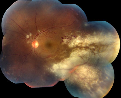
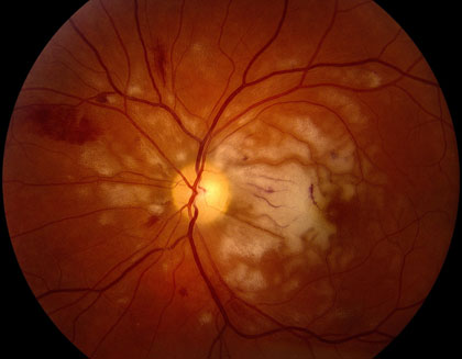
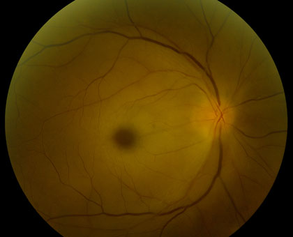
Retinal Artery Occlusions can be identified through the examination of the retina with special lenses, used to view the back of the eye. However, examinations do not always provide accurate indicators of a blockage. While disruption of blood flow, via segmentation (“box-carring”), can occur within seconds of initial vision loss, examinations may show normal results in these early stages. However, in the minutes to follow, as the retina becomes swollen from lack of oxygen, areas of retinal whitening become visible. While swelling usually resolves in weeks to months, producing clean retinal examination results, the retina might be permanently damaged.

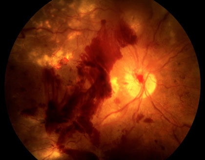
Fluorescein angiography can reveal a disruption of blood flow and can help identify the location of the blockage. Late complications, such as abnormal blood vessel growth (retinal neovascularization) can also be identified and may be used to guide treatment.

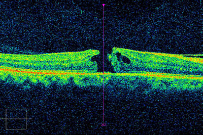
Optical coherence tomography (OCT) will show initial thickening of the retina, due to cellular edema followed by thinning of the retina, due to death of retinal tissue. Retinal thinning may be the only sign of a retinal artery occlusion, in the late stages of the disease.

Whose at risk?

Retinal Artery Occlusions are usually the result of an embolus (clot or plaque) from the carotid artery or the heart. An embolus, in either location, occludes blood flow to the retina. Because the retina is an extension of the brain, a retinal artery occlusion is considered a “stroke” of the eye. Approximately two-thirds of patients have underlying hypertension (high blood pressure) and one-fourth of patients will be found to have some preexisting disease. These diseases might include, significant carotid artery disease, characterized by the presence of plaque with a narrowing of the arterial lumen, cardiac valvular disease, or diabetes.

Some patients have one of the above diseases, combined with another underlying disorder. An extensive evaluation by an internal medicine physician may be required, using the carotid artery doppler ultrasound technology and echocardiography. Blood tests for clotting disorders (e.g., Protein S or C deficiency), elevated lipids (e.g., cholesterol and triglycerides) and inflammation (e.g., erythrocyte sedimentation rate and C-reacitve protein levels for Giant Cell Arteritis) may be required to identify risk factors in some patients.

Signs and Symptoms

In classic cases, Central Retinal Artery occlusion presents with a “cherry red spot”, caused by diffuse retinal whitening, which surrounds and, thus highlights, a central area of redness, called the fovea. The fovea is spared from the whitening due to the lack of retinal capillaries in the center of the retina. Branch retinal artery occlusion presents with a segment of retinal whitening, which extends from a blood vessel branch point. Symptoms of CRAO usually include severe vision loss; in fact, most patients can barely count the fingers in front of their face or see light, through the affected eye. Symptoms of BRAO, on the other hand, cover a broad range. In small peripheral occlusions, there may be zero central vision loss but in more widespread cases, vision loss can be severe, located centrally and/or directly above or below the center. Both conditions may be preceded by episodes of transient visual loss, known as amaurosis fugax.

Treatment

There is a very short window of opportunity (approx 90 minutes) available for the treatment, after the onset of vision loss. However, later rescue has been reported. While no tried-and-true standard of care exists, a diversity of treatments has been utilized with some reported success. If an embolus is identified, decreasing the eye pressure or dilating the retinal vessels may disrupt the blockage and restore vision. Pressure can be decreased, by way of fluid removal, in a number of ways, including needle extraction, ocular massage or glaucoma drops. Dissolving a blood clot is another alternative. A drug called “TPA” has been administered for this purpose, via direct vessel injection or systemic injection. It should be noted, however, that there has been some reported risk of stroke with this procedure. Other affective treatment methods include, rupturing plaque with a YAG laser, breathing into a paper bag to increase CO2, vessel dilation, hyperbaric oxygen treatments and steroid use. Systematic disease treatments, such as a carotid endarterectomy, cardiac valve replacement, anti-hypertensive drug therapy, lipid lowering agents and anti-coagulants may also be suggested. If ocular complications, such as a vitreous hemorrhage or rubiosis, occur as a result of treatments, anti-VEGF drugs (eg, Avastin), laser surgery or vitrectomy surgery may be employed. New restorative treatments are now available and we specialize in those therapies here at the Retina Macula Institute.

Retinal Tear and Detachment

What is Retinal Tear and Detachment?

The retina is a sensory tissue lining the inner surface of the eye. Retinal detachment is a serious condition that occurs when the inner layer of the retina separates from the eye wall.

Incoming light from the outside world triggers nerve impulses in the retina, which send visual information to the brain. When the retina detaches, the nerve cells have reduced functionality, potentially causing blurriness and vision loss.

Retinal detachment requires immediate medical care. If treated early on, vision can be restored.

Diagnosis

To diagnose retinal detachment, your doctor will examine your eyes and ask you questions regarding any symptoms you may be experiencing.

If you have experienced symptoms of retinal detachment, your doctor will use a lighted magnifying tool called an ophthalmoscope to examine your retina. With this tool, your doctor can detect holes, tears, or a complete retinal detachment. Alternatively, an ultrasound may be used if the patient has a dense cataract, the eye is filled with blood, or if the patient is uncooperative. The underlying cause of one particular brand of detachment called, rhegmatogenous retinal detachment, is a retinal break. To detect a break, an examination of the retinal periphery might be recommended, using an indirect ophthalmoscope and sclera depression.

Who is at risk?

Retinal detachment often begins when the thick fluid that fills the center of the eye, called the vitreous gel, shrinks and separates from the retina. This sometimes-harmless phenomenon is referred to as a posterior vitreous detachment (PVD) and often occurs with age. In cases where the vitreous gel is strongly attached to the retina, PVD’s can occasionally cause more severe damage. When the vitreous gel shrinks, it will create so much tension that the retina might tear. This tear allows fluid to collect under the retina, sometimes causing the retina to detach completely. The 3 forms of retinal detachment include: rhegmatogenous, tractional and exudative.

Other causes of retinal detachment include eye or head injury, nearsightedness (myopia), eye disease, and conditions such as diabetes.

Unfortunately, most cases of retinal detachment cannot be prevented. But you can reduce your risk of retinal detachment with regular visits to your eye doctor and by wearing protective helmets and eyeglasses. Also, if you suspect that you might have diabetes or have been diagnosed previously, proper treatment is crucial in risk reduction.

Signs and Symptoms

retinal-detachment_4203.jpg

Many people experience symptoms of a posterior vitreous detachment before they have symptoms of retinal detachment. When the vitreous gel shrinks and separates from the retina, it causes floaters and flashes. Floaters are spots, specks, and lines that float across your field of vision. Flashes are brief sparkles or lightning streaks that are most easily seen when your eyes are closed. Floaters and flashes do not always mean that you will have a retinal detachment but they may be warning signs. It is best to be seen by a doctor if you experience an increase in these symptoms. Sometimes a retinal detachment occurs without warning. The first sign of detachment may be a permanent shadow across a portion of your vision field. Alternatively, you may experience a new and sudden loss of peripheral vision, which worsens over time.

Treatment

Retinal detachment requires immediate care. Without treatment, vision loss can progress, often resulting in blindness within hours or days. Surgery is the only way to reattach the retina. In most cases, surgery can restore a good amount of vision. There are several surgical methods, including the use of lasers or a freezing probe to seal the tear in the retina. In complex cases, a vitrectomy, characterized by the removal of the vitreous gel, may be required. Another method uses the injection of a gas or oil bubble to hold the retina in place during healing. A silicone band or scleral buckle may also be required. Patients must pay careful attention to post-operative instructions in order to increase their chances of success.

The goal of treatment is to reduce the risk of further vision loss while reversing current visual impairment. In complex or advanced cases, vision restoration may not be possible but the eye, itself, might be saved with surgery. Newer restorative treatments are now available for patients with complex retinal detachments. We specialize in these therapies here at the Retina Macula Institute.

Retinal Vein Occlusion

What is Retinal Vein Occlusion?

Second only to diabetic retinopathy, Retinal Vein Occlusions remains one of the leading causes of blindness, as a result of retinal vascular disease. This is caused by circulatory blockage of a retinal vein by an adjacent blood vessel and can take two different forms. Central Retinal Vein Occlusion (CRVO) occurs at the point where the vein enters the eye and, therefore, has a more overwhelming affect on vision. Branch Retinal Vein Occlusion (BRVO) affects only a segment of the circulation and has less of an influence on vision

Diagnosis

CRVO_420.jpg

The diagnosis of a retinal vein occlusion is made through the examination of the retina, at the back of the eye, using special lenses and a microscope. Early on, specifically patterned patches of blood and fluid may be detected. However, this signifier often fades with time, making diagnosis more difficult. The less obvious signs of a vein occlusion may be unmasked with the aid of fluorescein angiography, during which dye is injected into to the blood stream. This allows circulation of the retina to be measured. Another advanced method, called Optical Coherence Tomography (OCT) might be used to measure retinal swelling and/or to gauge the response to treatment. Furthermore, Electroretinography, can be used to measure the eye’s electrical response to light flashes, at a cellular level. In some cases, this can determine the prognosis and guide treatment.

Who is at risk?

Retinal vein occlusions appear to affect males and females equally. While the occurrence is higher in people over the age of 50, there are other significant risk factors, which may be prevented. Cardiovascular disease and diabetes both increase the risk of occlusions. High blood pressure and smoking triples the likelihood of disease, the latter inhibiting blood flow through hardening of the arteries. While you can’t change your age or family history, proper diet and a healthy active lifestyle can make great strides toward prevention. It is important to talk with both your medical and eye doctor to work on reducing personal risk factors.

Signs and Symptoms

Signs of a retinal vein occlusion may include the dilation of retinal veins due to patches of swelling (edema) and/or blood (a hemorrhage) in the retina. Symptoms often include transient blurring of vision, which may last for hours or days. These episodes are usually followed by the onset of diffuse or segmental vision loss, which can be mild or severe. In a non-ischemic vein occlusion, where a partial blockage is present, vision loss may be mild. In an ischemic vein occlusion vision loss is often more severe and complications more common. Here, ischemia refers to a decrease in blood supply to the eye. It is possible for a patient, suffering from a peripheral vein occulsion, to be asymptomatic.

Treatment

Traditional treatment for vein occlusions includes the thermal laser method or simple observation (no treatment). Observation is indicated for patients who have non-ischemic (partial) vein occlusions and good vision (e.g., 20/30 or better). However, these patients should still be monitored closely; progressive vision loss may occur, along with other complications, which might prompt treatment. Laser may be used to improve vision for patients with significant macular edema or to reduce the risk of blindness due to abnormal blood vessel growth in the front or back of the eye. New treatments, currently being researched, include the intraocular injection of medications, such as anti-VEGF drugs (e.g., Avastin, Lucentis or Macugen) and steroids (e.g., triamcinolone acetate) to stop abnormal blood vessel growth and leakage. Surgical treatment is less common with the dawn of these new medications but it still exist. These surgeries may include a vitrectomy, with membrane peeling and laser therapy. For Branch Vein Occlusion, a sheathotomy might be performed or, for Central Vein Occlusion, a radial optic neurotomy. Newer treatments are now available that have proven successful in restoring vision; we specialize in these therapies here at the Retina Macula Institute.

Uveitis

What is Uveitis?

uveitis_420.jpg

Uveitis (pronounced you-vee-EYE-tis) is an inflammation of the tissues that occupy the middle layers of the eye. These layers are called the uveal tract or uvea. The uvea includes the iris (colored part of the eye), choroid (a thin membrane containing many blood vessels) and ciliary body (the part of the eye that joins these together).The uvea is a critical structure for vision because it contains the blood vessels that supply oxygen and nutrients to the critical parts of the eye. While uveitis is uncommon, affecting about 1 in 1000 adults and 1 in 2000 children, it can cause serious vision loss or blindness if not diagnosed early and treated aggressively.

Diagnosis

In some cases, a diagnosis can be made, based on patient history and on an eye examination, alone. Changes in vision are not always indicative of a clear diagnosis, so an evaluation of potential underlying causes is required. This may include a work-up, searching for inflammation, infection or infiltration (cancer). Laboratory tests including blood work, a TB skin test and X-rays are usually the starting point. Because uveitis may originate elsewhere in the body, your primary care doctor or a specialist may be required to conduct a complete medical exam.

Signs and Symptoms

The main signs of uveitis include eye redness, abnormal eye pressure levels, a cloudy film in the eye from inflammatory cells, retinal swelling and blood vessel leakage in the back of the eye. Symptoms include light sensitivity, eye pain, blurred vision, and floaters.

Uveitis may develop rapidly, and it is very important that you see your eye doctor (ophthalmologist or optometrist) for a complete eye examination if you develop any of the above symptoms. Be especially wary if a painful red eye does not clear up quickly. Left untreated, uveitis may permanently damage your vision.

Diagnosis

Uveitis has many potential causes, including viral infections, fungus, bacteria, parasites, peripheral inflammatory disease, or eye trauma. There are four types of uveitis and tests target specific diagnoses: Iritis is the most common form of uveitis. It affects the iris and is often associated with autoimmune disorders such as rheumatoid arthritis. Iritis may develop suddenly and may last up to eight weeks, regardless of treatment. Cyclitis is an inflammation of the middle portion of the eye and may affect the muscle responsible for focusing the lens. Cyclitis may develop suddenly and last for several months. Retinitis affects the back of the eye. It can progress rapidly, making it difficult to treat. Viruses, such as shingles or herpes, may cause retinitis. It can also be caused by bacterial infections, such as syphilis or toxoplasmosis. Choroiditis is marked by inflammation of the tissue layer, beneath the retina. It may also be caused by an infection such as tuberculosis or an autoimmune disease like rheumatoid arthritis or lupus. In a large number of cases, the cause of uveitis is not known. Reducing stress can be a helpful preventative measure.

Treatment

Because uveitis is serious, treatment needs to begin immediately. If infection is not the cause, your ophthalmologist may prescribe steroid eye drops, in order to reduce swelling, in combination with pain relievers. Antibiotics are used in patients with infectious uveitis and dark glasses are issued to ease light sensitivity. Complications of uveitis may include glaucoma, cataracts, retinal fluid build-up, vision loss and abnormal blood vessel growth – the latter of which might interfere with vision. Early diagnosis and treatment is critical. Newer treatments are now available that can restore vision in some patients with severe uveitis; we specialize in these therapies here at the Retina Macula Institute.

Retinitis Pigmentosa and other Retinal Degenerations

Retinitis Pigmentosa (RP) is an inherited disorder that affects the light-sensitive cells in the eyes, called the photoreceptors. The photoreceptors are made up of rods and cones. The rods are the cells that are most sensitive to dim light and, therefore, are the primary victim of RP. Cones are responsible for color and central fine-detail vision and are affected secondarily. RP can be caused by a number of disorders. Different genes may cause different degrees of vision loss and the onset and severity can vary as well.
Signs and Symptoms

Most patients with RP will experience night vision loss as an initial symptom. They will struggle to see stars and will have trouble driving at night. The rods are responsible for this type of vision and because they are located in the periphery, a patient might eventually experience a loss of side vision altogether (“tunnel vision”). Patients might test 20/20 but they will be considered legally blind because of the degree to which their peripheral vision is compromised. Eventually the central vision might also deteriorate; the signs of central vision loss can be detected by your eye doctor and include retinal pigmentation with the appearance of “bone-spicule” deposits. This will occur when the arteries that carry oxygen to the retina narrow and the optic nerve – containing the cells that carry the signals to the brain – become pale. Other signs can be detected with more specific tests. For instance, the visual field test might show significant contraction and, when less than 20 degrees of the field remains, the patient is considered legally blind. The elctroretinogram (ERG) measures the electrical signal of the retinal cells in response to light; in advanced cases this reaction is suppressed or perhaps absent altogether. In patients with RP, an ERG will show a loss of the “scotpac signal” or the dark-adapted response of the retina. Other tests like Fluorescein Angiography and Optical Coherence Tomography can detect cell loss or “atrophy” found in RP patients, which might not have been spotted in a regular eye exam. Many patients with RP will develop cataracts, so your eye doctor should keep close watch for the symptoms. Other treatable problems, like Cystoid Macular Edema, might also be detected with these advanced tests. Recently, genetic testing for some forms of RP has been made possible; while the tests are currently used for diagnosis, they may ultimately be useful for treatment, as well.

What is the treatment for Retinitis Pigmentosa?

While there is no cure for RP, there are treatments that can help slow vision loss. Dicosahexaenoic Acid (DHA) and the retinal palmitate form of vitamin A have both been shown to slow visional field loss. Diets enriched in Omega 3 fatty acids have also been shown to slow vision loss. Vitamin E, however, – in doses of 400 IU – has been found to accelerate the progression of RP. If started by the age of 30 and, with the appropriate vitamin regimen and diet, a patient can add as many as 20 years of functional vision to their lives. While there are no reported instances of toxicity in healthy adults taking 15,000 IU of vitamin A palmitate daily, people are advised to have a liver function test completed and their vitamin A blood levels measured before they begin the regimen. These tests should be revisited annually, thereafter. Also, because of the potential for birth defects, women who are pregnant or planning to become pregnant are not advised to take vitamin A palmitate in this dosage. Bone density in women may also be an issue.

For patients with CME, anti-inflammatory drops/injections and a diuretic, called Acetazolamide, may be effective. PSC cataract is amenable to surgery. Clinical trials with stem cells and growth factor replacement are currently underway and the specialists at RMI have helped pioneer some of the treatments. We keep our patients up-to-date about the latest trials and treatment options, so to help them maximize their visual potential.